Dissecting sheep brain
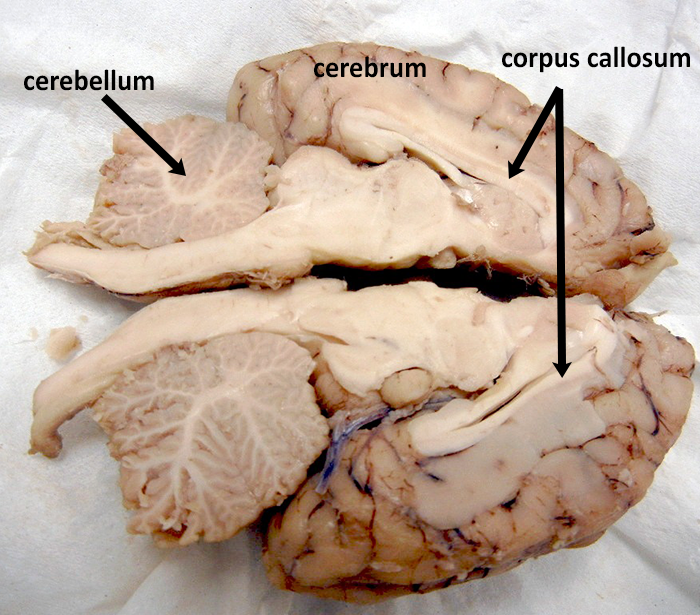
Dissecting Sheep Brain. Use a knife or a scalpel to cut the specimen along the longitudinal fissure. This will allow you to separate the brain into the left and right hemispheres. Recorded at glen oaks community college centreville michigan by dr ren allen hartung. Closely examine this sheep organ to learn about structures of the brain such as the cerebellum cranial nerve and so much more.
 Ihs Synapsis Sheep Brain Dissection The Irvington Voice From ihsvoice.com
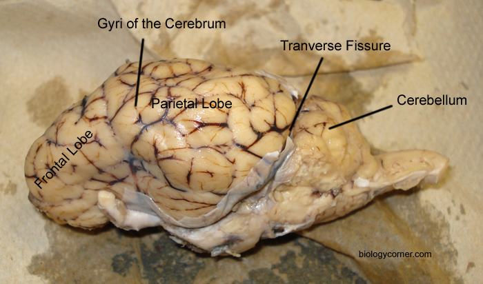
Take special note of the pituitary gland and the optic chiasma. Muscles other nerves and fatty. Examine the ventral surface of the sheep brain. Place the brain with the curved top side of the cerebrum facing up. Sheep brain dissection guide and steps when completing this procedure. Identify the cerebrum and the medial longitudinal fissure which separates the right and left hemispheres of the cerebral cortex.
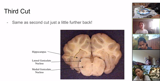
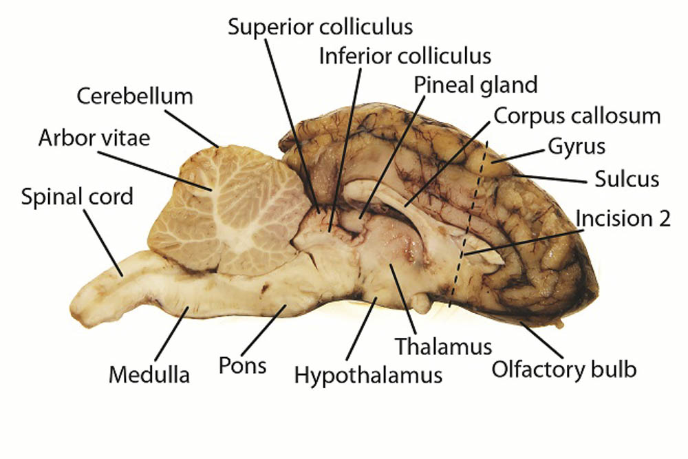
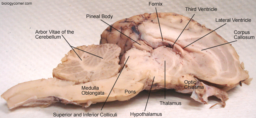
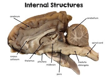
Lay one side of the brain on your tray to locate the structures visible on the inside.
You should also cut through the cerebellum. Use a knife or a scalpel to cut the specimen along the longitudinal fissure. The sheep brain is enclosed in a tough outer covering called the dura mater. Observe the structure of the of the sheep brain. An image of the brain is included to help students find the structures. Locate the four lobes of the.
 Source: learning-center.homesciencetools.com
Examine the ventral surface of the sheep brain. The next several steps will view this surface of the brain. Separate the two halves of the brain and lay them with the inside facing up. Also the sheep brain is oriented anterior to posterior whereas the human brain is superior to inferior. A pair of olfactory bulbs may be seen one under each lobe of the frontal cortex.
 Source: brainu.org
The sheep has a smaller cerebrum. The sheep brain is quite similar to the human brain except for proportion. Take special note of the pituitary gland and the optic chiasma. Examining the internal sheep brain. Observe the structure of the of the sheep brain.
 Source: ihsvoice.com
You will need. An image of the brain is included to help students find the structures. Place the brain with the curved top side of the cerebrum facing up. Lay one side of the brain on your tray to locate the structures visible on the inside. Also the sheep brain is oriented anterior to posterior whereas the human brain is superior to inferior.
 Source: pinterest.com
A preserved sheep brain specimen a photographic sheep brain dissection guide a 22 scalpel a magnifying glass and a dissecting tray. Several important parts of the visual system are visible in the ventral view of the brain. As students dissect a sheep brain they ll even gain a better understanding of. Brain with dura mater intact. Identify the cerebrum and the medial longitudinal fissure which separates the right and left hemispheres of the cerebral cortex.
 Source: biologycorner.com
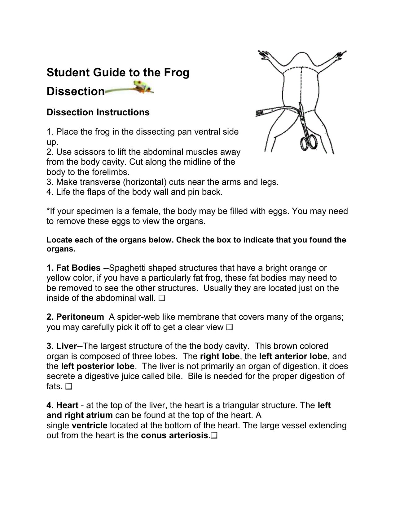
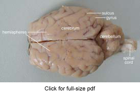
Place the brain on a dissection tray dorsal side up. Dissection and description of the anatomy of the sheep brain. Dissection guide with instructions for dissecting a sheep brain. The sheep brain is quite similar to the human brain except for proportion. You will need.
 Source: biologycorner.com
Checkboxes are used to keep track of progress and each structure that can be found is described with its location in relation to other structures. You should also cut through the cerebellum. The sheep brain is enclosed in a tough outer covering called the dura mater. Identify the cerebrum and the medial longitudinal fissure which separates the right and left hemispheres of the cerebral cortex. Examine the ventral surface of the sheep brain.
 Source: brains-explained.com
Identify the cerebrum and the medial longitudinal fissure which separates the right and left hemispheres of the cerebral cortex. Closely examine this sheep organ to learn about structures of the brain such as the cerebellum cranial nerve and so much more. Brain with dura mater intact. Checkboxes are used to keep track of progress and each structure that can be found is described with its location in relation to other structures. Lay one side of the brain on your tray to locate the structures visible on the inside.
 Source: quizlet.com
A pair of olfactory bulbs may be seen one under each lobe of the frontal cortex. Use a scalpel or sharp thin knife to slice through the brain along the center line starting at the cerebrum and going down through the cerebellum spinal cord medulla and pons. Examining the internal sheep brain. Examine the ventral surface of the sheep brain. Checkboxes are used to keep track of progress and each structure that can be found is described with its location in relation to other structures.
 Source: courses.lumenlearning.com
Brain with dura mater intact. A pair of olfactory bulbs may be seen one under each lobe of the frontal cortex. An image of the brain is included to help students find the structures. Observe the structure of the of the sheep brain. Use a knife or a scalpel to cut the specimen along the longitudinal fissure.
 Source: biology4friends.org
Sheep brain dissection guide and steps when completing this procedure. Sheep brain dissection guide 3. Lay one side of the brain on your tray to locate the structures visible on the inside. Locate the four lobes of the. This will allow you to separate the brain into the left and right hemispheres.
 Source: biologycorner.com
As students dissect a sheep brain they ll even gain a better understanding of. Checkboxes are used to keep track of progress and each structure that can be found is described with its location in relation to other structures. Locate the four lobes of the. You will need. The tough outer covering of the sheep brain is the dura mater one of three meninges membranes that cover the brain.
 Source: youtube.com
The sheep brain is quite similar to the human brain except for proportion. Locate the four lobes of the. Sheep brain dissection guide and steps when completing this procedure. The next several steps will view this surface of the brain. Place the brain on a dissection tray dorsal side up.
 Source: youtube.com
These two structures will likely be pulled off when you remove the dura mater. As students dissect a sheep brain they ll even gain a better understanding of. Dissection guide with instructions for dissecting a sheep brain. The next several steps will view this surface of the brain. Recorded at glen oaks community college centreville michigan by dr ren allen hartung.
 Source: teacherspayteachers.com
An image of the brain is included to help students find the structures. These two structures will likely be pulled off when you remove the dura mater. Muscles other nerves and fatty. The next several steps will view this surface of the brain. You will need.
 Source: learning-center.homesciencetools.com
The tough outer covering of the sheep brain is the dura mater one of three meninges membranes that cover the brain. Observe the structure of the of the sheep brain. A pair of olfactory bulbs may be seen one under each lobe of the frontal cortex. Several important parts of the visual system are visible in the ventral view of the brain. Place the brain on a dissection tray dorsal side up.
If you find this site good, please support us by sharing this posts to your preference social media accounts like Facebook, Instagram and so on or you can also save this blog page with the title dissecting sheep brain by using Ctrl + D for devices a laptop with a Windows operating system or Command + D for laptops with an Apple operating system. If you use a smartphone, you can also use the drawer menu of the browser you are using. Whether it’s a Windows, Mac, iOS or Android operating system, you will still be able to bookmark this website.

