Frog internal anatomy labeled
Frog Internal Anatomy Labeled. We will also find out that some of the structures of the frog will explain the living characteristics and tendencies of the frog. As members of the class amphibia frogs may live some of their adult lives on land but they must return to water to reproduce. Internal nares nostrils breathing connect to lungs. Tube leading to the lungs.
 Frog Dissection Carolina Com From carolina.com
Frog internal anatomy dissection instructions 1. The first organ system you ll find is the digestive system. Koenig updated may 11 2011 3 08 pm this read color label worksheet has the same anatomy on each side. Once it s time to open your frog the first cut will be through the ventral side to expose the internal organs. I tell students to start with the reading side but they will ignore you. Place the frog in the dissecting pan ventral side up.
Eggs are laid and fertilized in water.
Equalize pressure in inner ear. Place the frog in the dissecting pan ventral side up. Dorsal the back or upper surface of an organism ventral the stomach or lower surface of an organism anterior head end of an organism posterior tail end of an organism. By doing so we will study some relationships between the internal and external anatomy of the frog. Front attached aids in grabbing prey. Make transverse horizontal cuts near the arms and legs.
 Source: carolina.com
Frog internal anatomy label color posted may 11 2011 12 05 pm by ms. Use scissors to life the abdominal muscles away from the body cavity. Internal anatomy dissection instructions. Frog internal anatomy dissection instructions 1. You can click the image to magnify if you cannot see clearly.
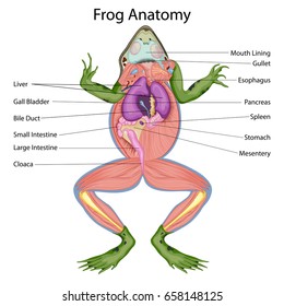 Source: shutterstock.com
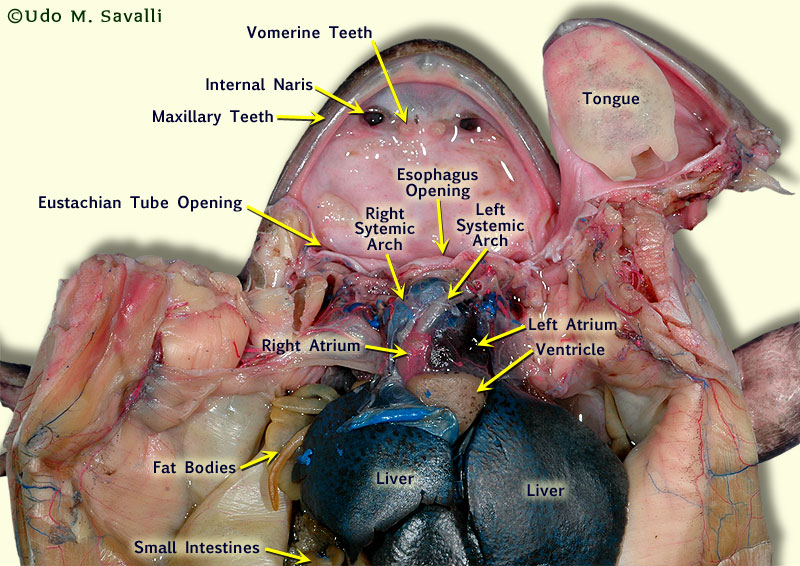
Front attached aids in grabbing prey. Internal anatomy dissection instructions. As members of the class amphibia frogs may live some of their adult lives on land but they must return to water to reproduce. Frog internal anatomy label color posted may 11 2011 12 05 pm by ms. Used for holding prey located at the roof of the mouth maxillary teeth.
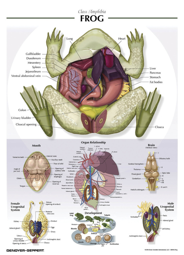 Source: schoolspecialty.com
By doing so we will study some relationships between the internal and external anatomy of the frog. Used for holding prey located at the roof of the mouth maxillary teeth. Internal anatomy dissection instructions. This image added by admin. Make transverse horizontal cuts near the arms and legs.
 Source: savalli.us
Make transverse horizontal cuts near the arms and legs. Use scissors to life the abdominal muscles away from the body cavity. Place the frog in the dissecting pan ventral side up. Frog internal and external anatomy. Frog internal anatomy label color posted may 11 2011 12 05 pm by ms.
 Source: biologyjunction.com
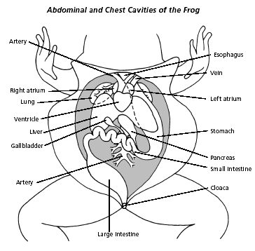
Two tympani or continue reading frog dissection. Life the flaps of the body wall and pin back. Internal nares nostrils breathing connect to lungs. Cut along the midline of the body from the pelvic to the pectoral girdle. One picture is simple and the other is more realistic but also low quality.
 Source: pinterest.com
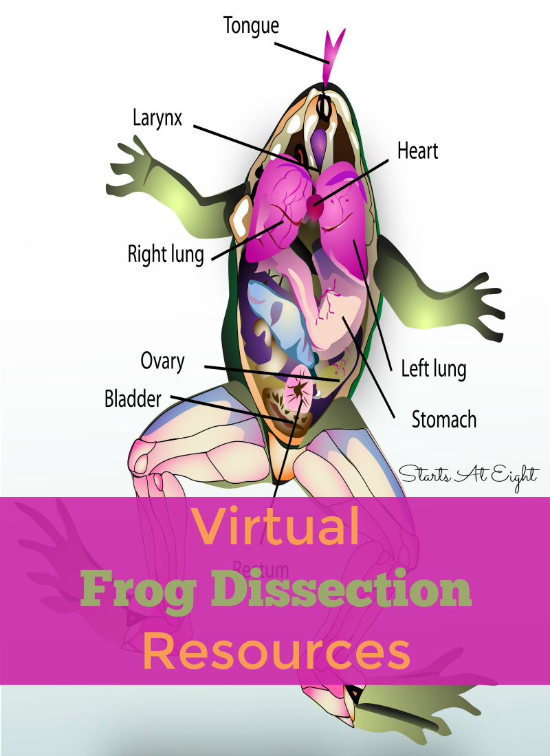
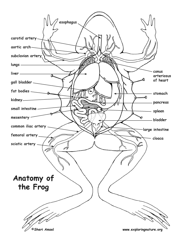
On the outside of the frog s head are two external nares or nostrils. Place the frog in the dissecting pan ventral side up. Labeled picture of a frog frog muscles labeled frog internal anatomy diagram labeled muscular internal part of wide collections of all kinds of labels pictures online. We think this is the most useful anatomy picture that you need. Place the frog in the dissecting pan ventral side up.
 Source: startsateight.com
Internal anatomy dissection instructions. Internal nares nostrils breathing connect to lungs. We will also find out that some of the structures of the frog will explain the living characteristics and tendencies of the frog. Make your work easier by using a label. Life the flaps of the body wall and pin back.
 Source: pinterest.com
One picture is simple and the other is more realistic but also low quality. Frog internal anatomy label color posted may 11 2011 12 05 pm by ms. You can click the image to magnify if you cannot see clearly. Two tympani or continue reading frog dissection. Make transverse horizontal cuts near the arms and legs.
 Source: exploringnature.org
Internal nares nostrils breathing connect to lungs. Modern biology holt background. Make your work easier by using a label. Frog internal and external anatomy. Two tympani or continue reading frog dissection.
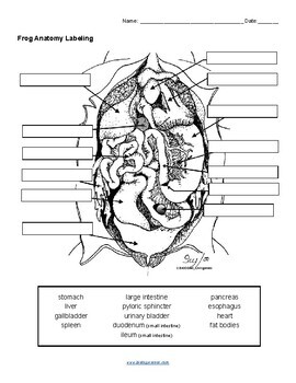 Source: teacherspayteachers.com
Cut along the midline of the body to the forelimbs. We will also find out that some of the structures of the frog will explain the living characteristics and tendencies of the frog. Labeled picture of a frog frog muscles labeled frog internal anatomy diagram labeled muscular internal part of wide collections of all kinds of labels pictures online. Two tympani or continue reading frog dissection. Dorsal the back or upper surface of an organism ventral the stomach or lower surface of an organism anterior head end of an organism posterior tail end of an organism.
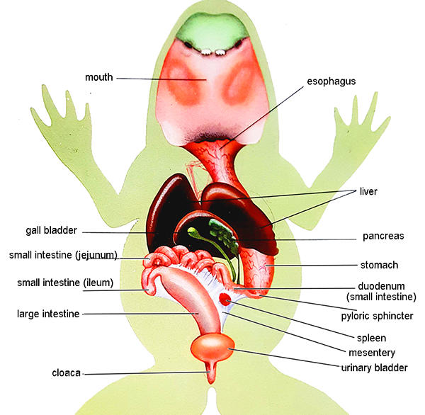 Source: biologycorner.com
Frog internal anatomy dissection instructions 1. Frog internal anatomy label color posted may 11 2011 12 05 pm by ms. Tube leading to the lungs. Labeled picture of a frog frog muscles labeled frog internal anatomy diagram labeled muscular internal part of wide collections of all kinds of labels pictures online. Equalize pressure in inner ear.
 Source: pinterest.com
Cut along the midline of the body from the pelvic to the pectoral girdle. Make your work easier by using a label. Koenig updated may 11 2011 3 08 pm this read color label worksheet has the same anatomy on each side. Place the frog in the dissecting pan ventral side up. By doing so we will study some relationships between the internal and external anatomy of the frog.
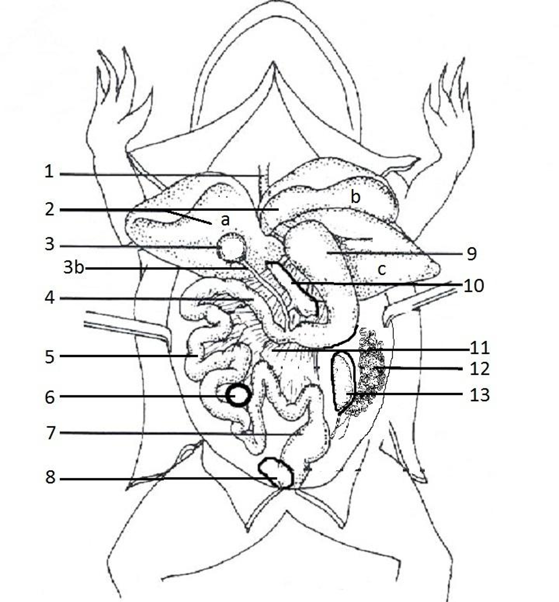 Source: biologycorner.com
Eggs are laid and fertilized in water. By doing so we will study some relationships between the internal and external anatomy of the frog. Used for holding prey located at the roof of the mouth maxillary teeth. Cut along the midline of the body from the pelvic to the pectoral girdle. Life the flaps of the body wall and pin back.
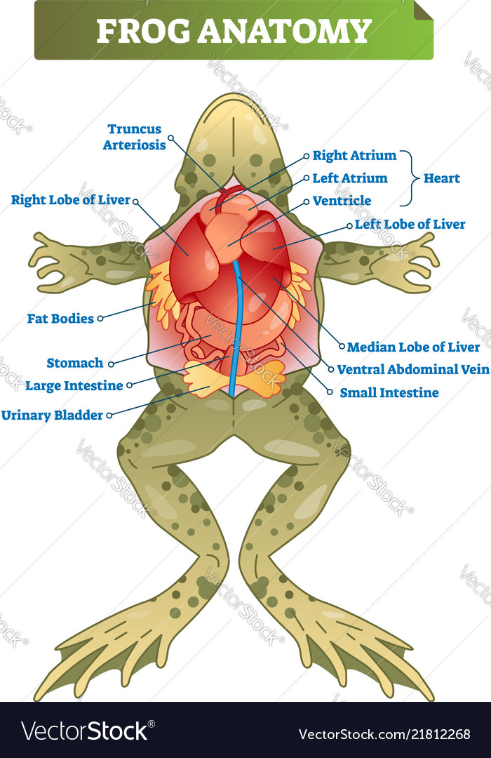 Source: vectorstock.com
Place the frog in the dissecting pan ventral side up. Dorsal the back or upper surface of an organism ventral the stomach or lower surface of an organism anterior head end of an organism posterior tail end of an organism. One picture is simple and the other is more realistic but also low quality. Used for holding prey located around the edge of the mouth. Used for holding prey located at the roof of the mouth maxillary teeth.
 Source: 123rf.com
We will also find out that some of the structures of the frog will explain the living characteristics and tendencies of the frog. I tell students to start with the reading side but they will ignore you. On the outside of the frog s head are two external nares or nostrils. We think this is the most useful anatomy picture that you need. Koenig updated may 11 2011 3 08 pm this read color label worksheet has the same anatomy on each side.
If you find this site adventageous, please support us by sharing this posts to your preference social media accounts like Facebook, Instagram and so on or you can also save this blog page with the title frog internal anatomy labeled by using Ctrl + D for devices a laptop with a Windows operating system or Command + D for laptops with an Apple operating system. If you use a smartphone, you can also use the drawer menu of the browser you are using. Whether it’s a Windows, Mac, iOS or Android operating system, you will still be able to bookmark this website.
