Onion root tip slide
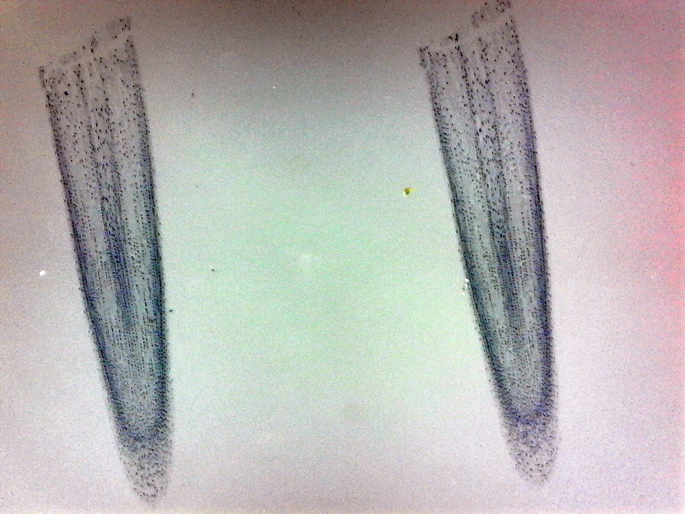
Onion Root Tip Slide. Add 2 3 drops of acetocarmine stain to the slide. Prepared microscope slide of a whitefish embryo. In order to examine cells in the tip of an onion root a thin slice of the root is placed onto a microscope slide and stained so the chromosomes will be visible. If you need more cells.

Discard the rest of the root. Prepared microscope slide of a whitefish embryo. The cells you ll be looking at in this activity were photographed with a light microsope and then digitized so you can see them on the computer. If you need more cells. You don t want to cook either your fingers or the root. This preparation of onion root tip cells is now ready for the study of mitosis.
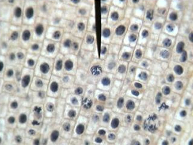
When observing the onion root tip cells for the stage of prophase the cells took on a brick like structure and within the cells small dots the nuclei can be seen.
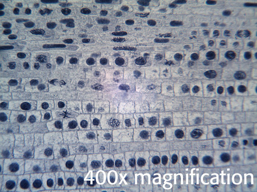
Under high power 400x move and focus the slide until the field of view is filled with as many cells in various phases of the cell cycle as possible. When observing the onion root tip cells for the stage of prophase the cells took on a brick like structure and within the cells small dots the nuclei can be seen. Observe and study mitosis by placing the slide under the compound microscope. The onion root tip cells slide is now prepared and ready to be examined for different stages of mitosis. Begin counting the cells one column at a time from left to right. This preparation of onion root tip cells is now ready for the study of mitosis.

Do not let the root dry out. Begin counting the cells one column at a time from left to right. Do not allow the slide to get hot to the touch. You will need to count at least 100 cells. Place the cut tip on a clean microscope slide.
 Source: southernbiological.com
Under high power 400x move and focus the slide until the field of view is filled with as many cells in various phases of the cell cycle as possible. Place the slide under the compound microscope and observe the different stages of mitosis. You don t want to cook either your fingers or the root. You will need to count at least 100 cells. Various stages of mitosis are prophase metaphase anaphase and telophase.
 Source: youtube.com
Prepared microscope slide of a whitefish embryo. An onion root tip is a rapidly growing part of the onion and thus many cells will be in different stages of mitosis. Various stages of mitosis are prophase metaphase anaphase and telophase. In order to examine cells in the tip of an onion root a thin slice of the root is placed onto a microscope slide and stained so the chromosomes will be visible. In one particular cell s nucleus the chromatin has condensed so.
 Source: amazon.com
Place the slide under the compound microscope and observe the different stages of mitosis. Add 2 3 drops of acetocarmine stain to the slide. Simulator procedure as performed through the online labs. In order to examine cells in the tip of an onion root a thin slice of the root is placed onto a microscope slide and stained so the chromosomes will be visible. Mitosis in onion root tips 1 mitosis in onion root tips 2 resting cell 3 early prophase 4 late prophase 5 metaphase 6 early anaphase 7 late anaphase 8 telophase 9 daughter cells 10 whitefish mitosis 11 interphase 12 prophase 13 metaphase 14 anaphase 15 telophase 16 cytokinesis to interphase.
 Source: pinterest.com
Discard the rest of the root. Do not let the root dry out. This preparation of onion root tip cells is now ready for the study of mitosis. Place the cut tip on a clean microscope slide. Various stages of mitosis are prophase metaphase anaphase and telophase.
Source: enasco.com
Place the cut tip on a clean microscope slide. Prepared microscope slide of an onion root tip. Focus as desired to obtain a distinct and clear image. Prepared microscope slide of a whitefish embryo. Do not let the root dry out.
 Source: instruction.greenriver.edu
Various stages of mitosis are prophase metaphase anaphase and telophase. An onion root tip is a rapidly growing part of the onion and thus many cells will be in different stages of mitosis. In order to examine cells in the tip of an onion root a thin slice of the root is placed onto a microscope slide and stained so the chromosomes will be visible. In one particular cell s nucleus the chromatin has condensed so. You will need to count at least 100 cells.
 Source: sciencegeek.net
Observe and study mitosis by placing the slide under the compound microscope. Do not let the root dry out. Place an onion root tip slide on the microscope stage. When observing the onion root tip cells for the stage of prophase the cells took on a brick like structure and within the cells small dots the nuclei can be seen. The onion root tips can be prepared and squashed in a way that allows them to be flattened on a microscopic slide so that the chromosomes of individual cells can be observed easily.
 Source: homesciencetools.com
Add 2 3 drops of acetocarmine stain to the slide. When observing the onion root tip cells for the stage of prophase the cells took on a brick like structure and within the cells small dots the nuclei can be seen. An onion root tip is a rapidly growing part of the onion and thus many cells will be in different stages of mitosis. Under high power 400x move and focus the slide until the field of view is filled with as many cells in various phases of the cell cycle as possible. Add 2 3 drops of acetocarmine stain to the slide.
 Source: acornnaturalists.com
Begin counting the cells one column at a time from left to right. The onion root tip cells slide is now prepared and ready to be examined for different stages of mitosis. Begin counting the cells one column at a time from left to right. Add 2 3 drops of acetocarmine stain to the slide. Do not allow the slide to get hot to the touch.
 Source: sccollege.edu
Prepared microscope slide of an onion root tip. Observe and study mitosis by placing the slide under the compound microscope. Place the slide under the compound microscope and observe the different stages of mitosis. Do not allow the slide to get hot to the touch. The onion root tip cells slide is now prepared and ready to be examined for different stages of mitosis.
 Source: amazon.com
Focus as desired to obtain a distinct and clear image. Place the slide under the compound microscope and observe the different stages of mitosis. When observing the onion root tip cells for the stage of prophase the cells took on a brick like structure and within the cells small dots the nuclei can be seen. Prepared microscope slide of a whitefish embryo. Simulator procedure as performed through the online labs.
Source: enasco.com
When observing the onion root tip cells for the stage of prophase the cells took on a brick like structure and within the cells small dots the nuclei can be seen. Observe and study mitosis by placing the slide under the compound microscope. You don t want to cook either your fingers or the root. Place an onion root tip slide on the microscope stage. Do not let the root dry out.
 Source: biology.stackexchange.com
Do not allow the slide to get hot to the touch. Under high power 400x move and focus the slide until the field of view is filled with as many cells in various phases of the cell cycle as possible. Do not allow the slide to get hot to the touch. You don t want to cook either your fingers or the root. Mitosis in onion root tips 1 mitosis in onion root tips 2 resting cell 3 early prophase 4 late prophase 5 metaphase 6 early anaphase 7 late anaphase 8 telophase 9 daughter cells 10 whitefish mitosis 11 interphase 12 prophase 13 metaphase 14 anaphase 15 telophase 16 cytokinesis to interphase.
 Source: thoughtco.com
You don t want to cook either your fingers or the root. Warm the slide gently over the alcohol lamp for about one minute. You will need to count at least 100 cells. In one particular cell s nucleus the chromatin has condensed so. Do not let the root dry out.
If you find this site beneficial, please support us by sharing this posts to your preference social media accounts like Facebook, Instagram and so on or you can also save this blog page with the title onion root tip slide by using Ctrl + D for devices a laptop with a Windows operating system or Command + D for laptops with an Apple operating system. If you use a smartphone, you can also use the drawer menu of the browser you are using. Whether it’s a Windows, Mac, iOS or Android operating system, you will still be able to bookmark this website.

