Seminiferous tubules slide
Seminiferous Tubules Slide. The rete testis the area where tubules coalesce and connect to efferent ductules is shown as irregularly shaped lumens lined by a simple cuboidal epithelium. Myoid cells these cells have the characteristics of smooth muscle cells forming part of the fibrous envelope around the. Each seminiferous tuble is intricately coiled averaging about 0 2 mm diameter and 50 cm in length. Present on the right hand side of the tunica albuginea is the mediastinum a thickened portion of the tunica albuginea that contains the rete testis.
 Male Reproductive From ouhsc.edu
The sperm lineage of cells will be discussed later. Seminiferous tubules leydig cells. Accessory gland seminal. The germinal lining contains both sertoli cells and the developing spermatocytes. The tubule walls consist of a multilayered germinal. Testes are composed largely of seminiferous tubules coiled tubes the walls of which contain cells that produce sperm and are surrounded by a capsule the tunica albuginea.
The tubule walls consist of a multilayered germinal.
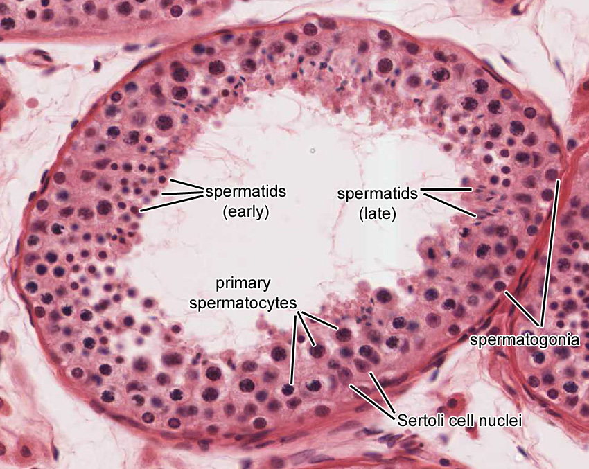
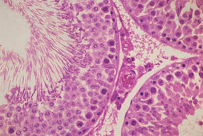
The seminiferous tubulewith its specialized germinal epitheliumis seen at higher power. Other articles where seminiferous tubule is discussed. The insides of the tubules are lined with seminiferous epithelium which consists of two general types of cells. The rete testis the area where tubules coalesce and connect to efferent ductules is shown as irregularly shaped lumens lined by a simple cuboidal epithelium. Spermatogenic cells and sertoli cells. Mature seminiferous tubules epon 100x.
 Source: histology.medicine.umich.edu
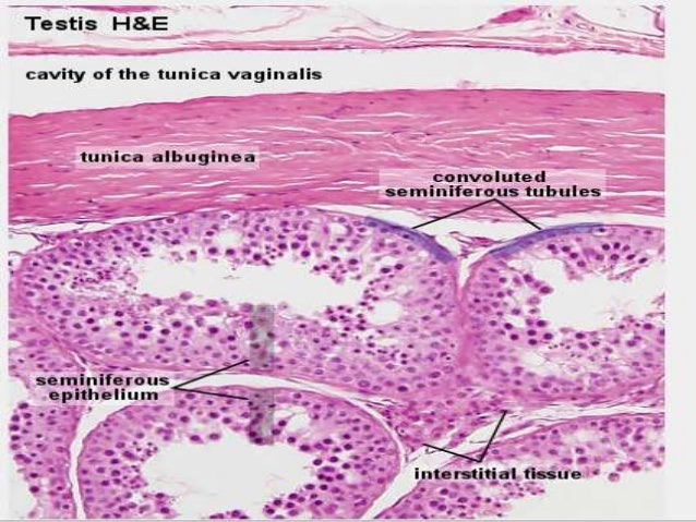
Seminiferous tubules leydig cells. Testes are composed largely of seminiferous tubules coiled tubes the walls of which contain cells that produce sperm and are surrounded by a capsule the tunica albuginea. Seminiferous tubules each testicular lobule contains 1 4 highly convoluted seminiferous tubules surrounded and supported by intertubular connective tissue. Spermatozoa is produced in the germinal epithelium of the seminiferous tubules and released into the lumina of these ducts. Present on the right hand side of the tunica albuginea is the mediastinum a thickened portion of the tunica albuginea that contains the rete testis.
 Source: instruction.cvhs.okstate.edu
The seminiferous tubulewith its specialized germinal epitheliumis seen at higher power. The seminiferous tubulewith its specialized germinal epitheliumis seen at higher power. The rete testis the area where tubules coalesce and connect to efferent ductules is shown as irregularly shaped lumens lined by a simple cuboidal epithelium. Various cell types are seen. These tubules are enclosed by a thick basal lamina and surrounded by 3 4 layers of smooth muscle cells or myoid cells.
 Source: medcell.med.yale.edu
Seminiferous tubules may constitute up to 90 percent of the testis. Seminiferous tubules the human testicular parenchyma contains several important structures. The insides of the tubules are lined with seminiferous epithelium which consists of two general types of cells. Each seminiferous tuble is intricately coiled averaging about 0 2 mm diameter and 50 cm in length. Male reproductive system testis thick connective tissue capsule connective tissue septa divide testis into 250 lobules tunica albuginea 1 seminiferous tubules each lobule contains 1 4 seminiferous tubules and interstitial connective tissue 2 rectus tubules 3 rete testis 4 efferent ductules 5 epididymis 6.
 Source: education.med.nyu.edu
Sertoli cells and spermatozoa are visible. Sertoli cells and spermatozoa are visible. Seminiferous tubules the human testicular parenchyma contains several important structures. Other articles where seminiferous tubule is discussed. Testis leydig cells.
 Source: medcell.med.yale.edu
Seminiferous tubules leydig cells. Myoid cells these cells have the characteristics of smooth muscle cells forming part of the fibrous envelope around the. The germinal lining contains both sertoli cells and the developing spermatocytes. Sertoli cells and spermatozoa are visible. The tubule walls consist of a multilayered germinal.
 Source: slideshare.net
The insides of the tubules are lined with seminiferous epithelium which consists of two general types of cells. Testis leydig cells. Seminiferous tubules leydig cells. This section features part of the testis on the lower portion of the slide. The seminiferous tubulewith its specialized germinal epitheliumis seen at higher power.
 Source: pinterest.com
These tubules are enclosed by a thick basal lamina and surrounded by 3 4 layers of smooth muscle cells or myoid cells. Male reproductive system testis thick connective tissue capsule connective tissue septa divide testis into 250 lobules tunica albuginea 1 seminiferous tubules each lobule contains 1 4 seminiferous tubules and interstitial connective tissue 2 rectus tubules 3 rete testis 4 efferent ductules 5 epididymis 6. Seminiferous tubules this is an audio histology slide. Seminiferous tubules leydig cells. Seminiferous tubules the human testicular parenchyma contains several important structures.
 Source: histology-world.com
Mature seminiferous tubules epon 100x. Individual tubules usually commence as free blind ends but neighbouring tubules may form anastomosing loops. The sperm lineage of cells will be discussed later. Testis leydig cells. Myoid cells these cells have the characteristics of smooth muscle cells forming part of the fibrous envelope around the.
 Source: ouhsc.edu
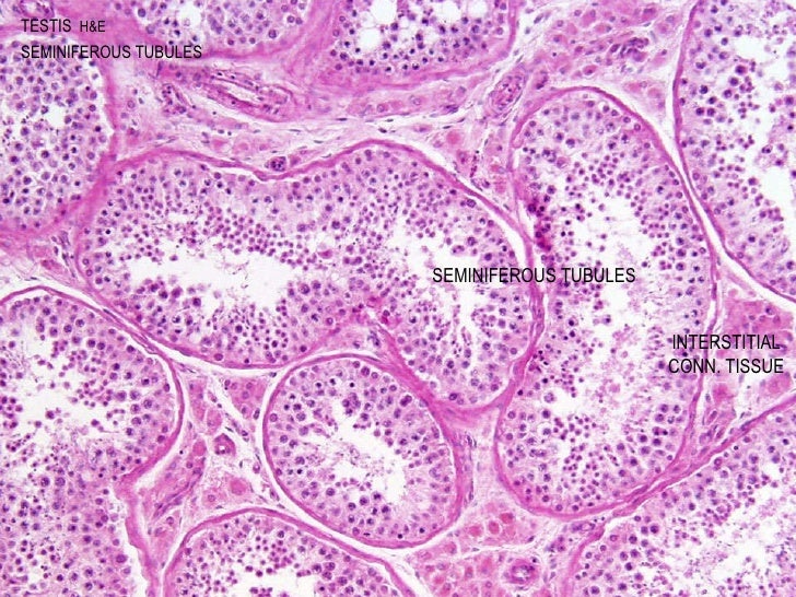
Between the tubules the interstitial cells of leydig and interstital connective tissue can be seen. Myoid cells these cells have the characteristics of smooth muscle cells forming part of the fibrous envelope around the. The tubule walls consist of a multilayered germinal. Accessory gland seminal. Seminiferous tubules may constitute up to 90 percent of the testis.
 Source: slideshare.net
The germinal lining contains both sertoli cells and the developing spermatocytes. Accessory gland seminal. These tubules are enclosed by a thick basal lamina and surrounded by 3 4 layers of smooth muscle cells or myoid cells. The rete testis the area where tubules coalesce and connect to efferent ductules is shown as irregularly shaped lumens lined by a simple cuboidal epithelium. Sertoli cells and spermatozoa are visible.
 Source: lab.anhb.uwa.edu.au
The tubule walls consist of a multilayered germinal. The insides of the tubules are lined with seminiferous epithelium which consists of two general types of cells. Myoid cells these cells have the characteristics of smooth muscle cells forming part of the fibrous envelope around the. Male reproductive system testis thick connective tissue capsule connective tissue septa divide testis into 250 lobules tunica albuginea 1 seminiferous tubules each lobule contains 1 4 seminiferous tubules and interstitial connective tissue 2 rectus tubules 3 rete testis 4 efferent ductules 5 epididymis 6. Mature seminiferous tubules epon 100x.
 Source: slideshare.net
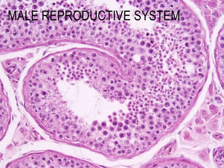
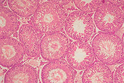
Sertoli cells and spermatozoa are visible. Each seminiferous tuble is intricately coiled averaging about 0 2 mm diameter and 50 cm in length. On these histology slides several seminiferous tubules are visible. The tubule walls consist of a multilayered germinal. Myoid cells these cells have the characteristics of smooth muscle cells forming part of the fibrous envelope around the.
 Source: histology-world.com
Note the seminiferous tubules and leydig cells. Accessory gland seminal. Seminiferous tubules may constitute up to 90 percent of the testis. The sperm lineage of cells will be discussed later. The germinal lining contains both sertoli cells and the developing spermatocytes.
 Source: ouhsc.edu
Testis leydig cells. The germinal lining contains both sertoli cells and the developing spermatocytes. Mature seminiferous tubules epon 100x. This section features part of the testis on the lower portion of the slide. The seminiferous tubulewith its specialized germinal epitheliumis seen at higher power.
 Source: ro.pinterest.com
Each seminiferous tuble is intricately coiled averaging about 0 2 mm diameter and 50 cm in length. Seminiferous tubules this is an audio histology slide. Accessory gland seminal. Testis leydig cells. On these histology slides several seminiferous tubules are visible.
If you find this site helpful, please support us by sharing this posts to your favorite social media accounts like Facebook, Instagram and so on or you can also save this blog page with the title seminiferous tubules slide by using Ctrl + D for devices a laptop with a Windows operating system or Command + D for laptops with an Apple operating system. If you use a smartphone, you can also use the drawer menu of the browser you are using. Whether it’s a Windows, Mac, iOS or Android operating system, you will still be able to bookmark this website.

