Step by step heart dissection
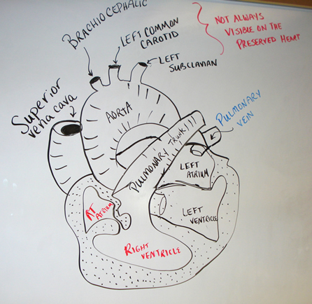
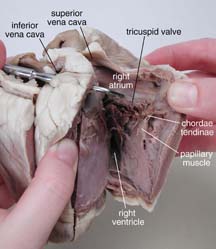
Step By Step Heart Dissection. Demonstration of a heart dissection including external and internal features the four main vessels connecting to the heart and an explanation of the blood. Observe the shape size and colour of the lungs and attached blood vessels leaving and entering the lungs. Don t be shy with the heart use your fingers to feel your way through the dissection. This is the opening to the superior vena cava which brings blood from the top half of the body to the right atrium the atria are the top chambers in the heart.
 Practical 1 Pig Heart Dissection From slideshare.net
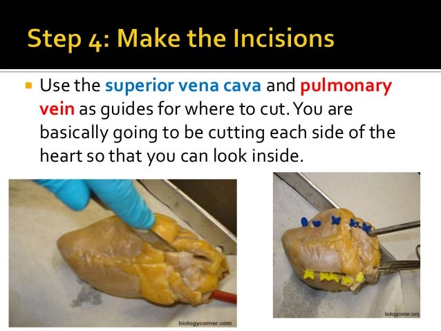
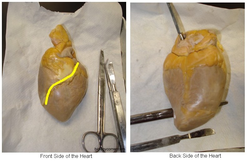
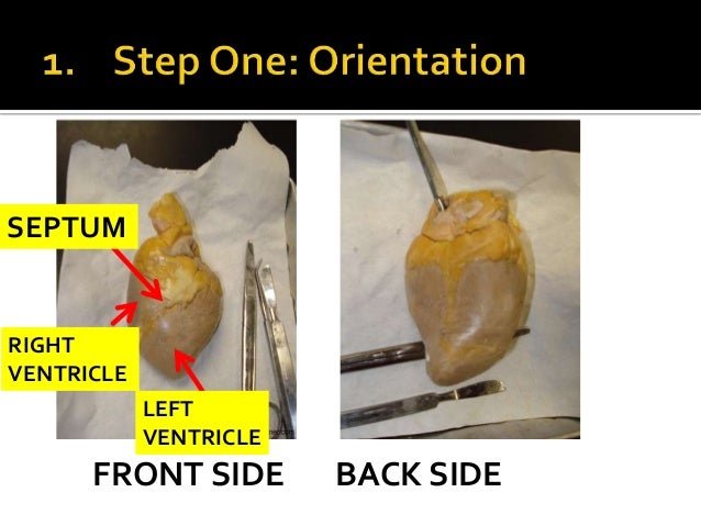
Find the line in the front of the heart and make an incision that isn t too deep only going through the top wall. This resource is designed for uk teachers. Investigating the external structure of the heart part 2. Before we start cutting make sure the heart s apex is facing you and the aorta is facing away from you. Identify the right and left sides of the heart on the left side of the heart make a cut from the pulmonary vein down to the left ventricle. This means that you really must experience the heart with your hands and feel your way to find the openings.
However you should watch the videos of the practical part 1.
Preview and details files included 1 rtf 2 mb. The fat is quite tuff so you need to apply a bit of force. This is an optional exercise so if you aren t able to do the practical or would rather not that s fine. If still attached identify the pleural membrane surrounding the lungs and the pericardium membrane surrounding the heart. Investigating the internal. Many people will be squeamish about this and because the heart is slippery it is easy to drop.
 Source: biologycorner.com
Brilliant sheet which guides you through a heart dissection. Lightly pull the two parts so it opens like a book and take a look inside. In the next few steps you ll have the opportunity to do a heart dissection at home. Identify the right and left sides of the heart on the left side of the heart make a cut from the pulmonary vein down to the left ventricle. 2 3 4 was a 3 month old p86 wild type c57bl 6 male.

The mouse imaged in figs. If using a pluck heart lung set arrange the pluck with heart on top on a dissecting board or tray. Brilliant sheet which guides you through a heart dissection. This is the opening to the superior vena cava which brings blood from the top half of the body to the right atrium the atria are the top chambers in the heart. This resource is designed for uk teachers.
 Source: biologycorner.com
However you should watch the videos of the practical part 1. The mouse imaged in figs. Find the line in the front of the heart and make an incision that isn t too deep only going through the top wall. The fat is quite tuff so you need to apply a bit of force. This is an optional exercise so if you aren t able to do the practical or would rather not that s fine.
 Source: tes.com
Preview and details files included 1 rtf 2 mb. Find the large opening at the top of the heart next to the right auricle. Don t be shy with the heart use your fingers to feel your way through the dissection. Observe the shape size and colour of the lungs and attached blood vessels leaving and entering the lungs. This is the opening to the superior vena cava which brings blood from the top half of the body to the right atrium the atria are the top chambers in the heart.
 Source: slideshare.net
The fat is quite tuff so you need to apply a bit of force. Many people will be squeamish about this and because the heart is slippery it is easy to drop. 2 3 4 was a 3 month old p86 wild type c57bl 6 male. This resource is designed for uk teachers. Brilliant sheet which guides you through a heart dissection.

However this particular dissection protocol can only be used on mice approximately p5 and above the older and larger the animal the easier the dissection. You should feel it open into the right atrium. The fat is quite tuff so you need to apply a bit of force. Observe the shape size and colour of the lungs and attached blood vessels leaving and entering the lungs. Mouse sensory neurons can be dissected and cultured as soon as they are formed in the embryo about embryonic day 13 e13.
 Source: youtube.com
If using a pluck heart lung set arrange the pluck with heart on top on a dissecting board or tray. The fat is quite tuff so you need to apply a bit of force. Before we start cutting make sure the heart s apex is facing you and the aorta is facing away from you. This is the opening to the superior vena cava which brings blood from the top half of the body to the right atrium the atria are the top chambers in the heart. Investigating the external structure of the heart part 2.
 Source: biologycorner.com
Don t be shy with the heart use your fingers to feel your way through the dissection. Before we start cutting make sure the heart s apex is facing you and the aorta is facing away from you. Mouse sensory neurons can be dissected and cultured as soon as they are formed in the embryo about embryonic day 13 e13. This resource is designed for uk teachers. 2 3 4 was a 3 month old p86 wild type c57bl 6 male.
 Source: learning-center.homesciencetools.com
If still attached identify the pleural membrane surrounding the lungs and the pericardium membrane surrounding the heart. If using a pluck heart lung set arrange the pluck with heart on top on a dissecting board or tray. Find the large opening at the top of the heart next to the right auricle. This is an optional exercise so if you aren t able to do the practical or would rather not that s fine. There are many ways you can cut your heart but the most informative will perhaps be directly down the interventricular sulcus.
 Source: carolina.com
There are many ways you can cut your heart but the most informative will perhaps be directly down the interventricular sulcus. If still attached identify the pleural membrane surrounding the lungs and the pericardium membrane surrounding the heart. Before we start cutting make sure the heart s apex is facing you and the aorta is facing away from you. This is the opening to the superior vena cava which brings blood from the top half of the body to the right atrium the atria are the top chambers in the heart. Observe the shape size and colour of the lungs and attached blood vessels leaving and entering the lungs.
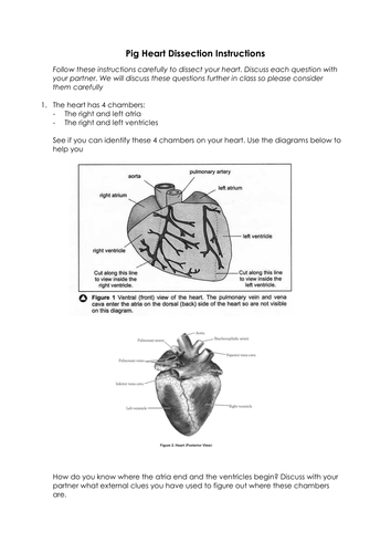
In the next few steps you ll have the opportunity to do a heart dissection at home. Investigating the internal. Don t be shy with the heart use your fingers to feel your way through the dissection. Investigating the external structure of the heart part 2. Lightly pull the two parts so it opens like a book and take a look inside.
 Source: pinterest.it
Investigating the internal. You should feel it open into the right atrium. Find the large opening at the top of the heart next to the right auricle. Lightly pull the two parts so it opens like a book and take a look inside. This is an optional exercise so if you aren t able to do the practical or would rather not that s fine.
 Source: slideshare.net
However you should watch the videos of the practical part 1. Before we start cutting make sure the heart s apex is facing you and the aorta is facing away from you. In the next few steps you ll have the opportunity to do a heart dissection at home. Identify the right and left sides of the heart on the left side of the heart make a cut from the pulmonary vein down to the left ventricle. Investigating the external structure of the heart part 2.
 Source: pinterest.com
You should feel it open into the right atrium. Mouse sensory neurons can be dissected and cultured as soon as they are formed in the embryo about embryonic day 13 e13. You should feel it open into the right atrium. If using a pluck heart lung set arrange the pluck with heart on top on a dissecting board or tray. Brilliant sheet which guides you through a heart dissection.
 Source: amazon.com
This is an optional exercise so if you aren t able to do the practical or would rather not that s fine. This is the opening to the superior vena cava which brings blood from the top half of the body to the right atrium the atria are the top chambers in the heart. In the next few steps you ll have the opportunity to do a heart dissection at home. This resource is designed for uk teachers. Observe the shape size and colour of the lungs and attached blood vessels leaving and entering the lungs.
If you find this site adventageous, please support us by sharing this posts to your preference social media accounts like Facebook, Instagram and so on or you can also bookmark this blog page with the title step by step heart dissection by using Ctrl + D for devices a laptop with a Windows operating system or Command + D for laptops with an Apple operating system. If you use a smartphone, you can also use the drawer menu of the browser you are using. Whether it’s a Windows, Mac, iOS or Android operating system, you will still be able to bookmark this website.

